|Articles|February 1, 2016
Biofilms & Catheters: The Mechanisms of Infection
Biofilms, or colonies of bacteria growing on surfaces and medical devices, can inflict intractable or recurring disease. During colonization, biofilms develop characteristics and behaviors more dangerous and powerful than those of planktonic (singleton) bacteria. In fact, these insidious microscopic collectives could be regarded as biological case studies in “strength in numbers” as they unify against external assault, resisting the host immune response as well as antimicrobials, and exact their high human and fiscal costs. Puzzlingly, although biofilms are a ubiquitous, well documented cause of infection, they receive only a modicum of the attention they clearly merit.
Advertisement
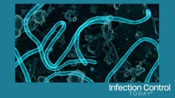
Biofilm on the septum/housing of a needleless connector. Image courtesy of Marcia Ryder, RN, MS, PhD
By Elizabeth Srejic
Biofilms, or colonies of bacteria growing on surfaces inside and outside of the body, can inflict intractable or recurring disease. During colonization, biofilms develop characteristics and behaviors more dangerous and powerful than those of planktonic (singleton) bacteria.1 In fact, these insidious microscopic collectives could be regarded as biological case studies in “strength in numbers” as they unify against external assault, resisting the host immune response as well as antimicrobials, and exact their high human and fiscal costs.2-3 Puzzlingly, although biofilms are a ubiquitous, well documented cause of infection, they receive only a modicum of the attention they clearly merit.
Biofilms can be particularly dangerous when they grow on medical devices inserted into the body.4 Specifically, biofilms present on intravascular catheters (IVCs) or central lines represent the most common cause of hospital-acquired septicemias: catheter-related bloodstream infections (CRBSIs).5 IVCs, which are often left in place for several weeks, provide a means to draw blood and administer medications and nutrition, but also furnish bacteria with an easily traversable superhighway leading directly into the bloodstream. And when microorganisms introduced from the skin of the patient at the catheter insertion site, from a contaminated catheter hub, or from hematogenous seeding of the device can attach to the device’s external surfaces and lumen (internal surfaces), infection becomes likely.6-7 Studies have shown that biofilms may form with-in three days after catheter insertion and tend to form on the external surface of catheters in place for less than 10 days; however, with in-creasing catheter duration (greater than or equal to 30 days), biofilms tend to form in the catheter lumen.8
“The introduction of a vascular catheter into the bloodstream triggers a host response,” says Marcia Ryder, RN, MS, PhD, a nationally and internationally recognized expert in the use and management of vascular access devices who has served as past-president of the Association for Vascular Access (AVA), past chair of the Scientific Research Council of the Association for Professionals in Infection Control and Epidemiology (APIC) and a former member of the Food and Drug Administration (FDA)'s Central Venous Catheter Working Group. “Plasma proteins, platelets and white blood cells adhere to the catheter forming a fibrinous layer that changes the surface properties for interaction with existing or arriving microorganisms. Bacteria are transferred to the external surface of the catheter from the patient’s resident skin flora, transient flora or caregiver’s hands during insertion of inadequately disinfected skin or with improper post-insertion maintenance techniques. Bacteria are transferred to the internal surface of the administration system that includes administration tubing, extension tubing, needleless connectors, injection ports, stopcocks, catheter hubs, and the catheter body by injecting through improperly disinfected access sites or touch contamination when separating connection points. Contaminated parenteral solutions and multi-dose vials continue to be a potential source of bacterial transmission.”
CRBSI is one of the most serious complications in hospitalized patients, leading to increased hospitalization, intensive care admissions, extensive antibiotic treatment, significant morbidity and d prolonged hospital stay and mortality.9 Each year in the United States, approximately 80,000 CRBSIs occur in intensive care unit (ICU) patients -- with an attributable cost of care estimated at $34,508 to $56,000 per episode -- and up to 250,000 CRBSIs occur throughout the healthcare system, accounting for a significant portion of HAIs.10-11
In spite of epidemiologic evidence associating biofilms and numerous infectious diseases, the exact mechanisms by which biofilm-associated microorganisms elicit disease are poorly understood.12 Currently, these mechanisms are thought to be detachment and embolization of cells or cell aggregates originating from the biofilm, endotoxin production, increased resistance to the host immune system, and creation of an antibiotic-resistant stronghold far more tenacious than planktonic (singleton) bacteria.13 This heightened protection is thought to arise at least in part from their secretion of a sticky extracellular polymeric matrix that spares the lower layers of bacteria thereby protecting the colony from complete eradication.14-15 Such tenacity can lead to chronic and recurring infections such as endocarditis, cystic fibrosis, cystitis, periodontitis, rhinosinusitis and osteomyelitis.16-17
“Self-preservation is a universal behavior among all living organisms that ensures survival,” Ryder says. “Microorganisms have exhibited extraordinary capacity to survive harsh environments over billions of years. As with animals and humans, safety and survival is optimized by cooperative responses of a group effort. Microorganisms utilize a ‘chemical and electrical intellect’ to communicate and cooperate in the establishment of a structured community on surfaces (both tissue and devices) with infrastructure to enable strategies of sustained living and protection from cellular (example, white blood cells) and chemical threats (example, antibiotics) in the surrounding biological environment. The self-produced structural component of the community is composed of a species-specific gelatinous type substance that is heterogeneous in oxygenation and nutrient availability. In the body, microorganisms involve host materials at the location of invasion (i.e. urine or blood proteins) in biofilm formation that adds protection. Further adaptation involves the up- and down-regulation of hundreds of cellular genes that alter metabolism and trans-form the organisms to a resistant survival mode.
“Microorganisms that contact any of the extra or intraluminal surfaces of any of these components produce attaching adhesins and in the presence of multiple organisms can form a biofilm either on the naked polymer of the device, or in conjunction with the proteinaceous conditioning layer or associated thrombus,” Ryder says. “The organisms divide exponentially and produce the embedding lubricious exopolymer matrix forming a colony. The biofilm may develop as patchy colonies or as a continuous layer. When the colony reaches a growth level that can no longer sustain growth, they begin the dissemination of organisms from the colony by enzymatic cleaving of clumps of biofilm or individual cells. Importantly, the embedding matrix is impermeable to immune cells resulting in premature release of endotoxins by white blood cells attempting phagocytosis resulting in an inflammatory response. Metabolically active cells at the surface of the biofilm may be transported away from the biofilm by high-flow shear stress, blood flow or intravenous infusion. Bacterial transfer into the bloodstream can be in considerable numbers depending on the biofilm burden on the device. Often overlooked in the dissemination of biofilm bacteria, particularly in the clump formation, are the deadly metastasizing infections including endocarditis, osteomyelitis, encephalitis, and abscess.”
“Vascular catheters are globally the most commonly used medical device in medical practice,” Ryder adds. “While these devices are often lifesaving and integral to a myriad of medical treatments, they also have significant risk for harm. As such, these devices represent a major patient safety concern and a focus of attention for infection control. Improvements have been made but we seem to have reached a plateau, at least in the United States0, in our efforts to achieve the expected reductions in catheter-related bloodstream infections. Our attention is now best directed to intense investigation of the science of prevention, diagnosis and treatment of biofilm related infectious diseases. Biofilm is the precursor of about 80 percent of infections including medical device and chronic infections. Our current approach to diagnosis and treatment of bio-film-related infections are ineffective as we continue to use a methodology specific to planktonic bacteria. Until the science develops to conquer the biofilm mode of infection, our best defense is to prevent the access of microorganisms to the surface(s) of the indwelling medical device. All forms of infusion therapy are technology dependent. Both research and development of new and existing technology and clinical efficacy of new devices should involve a rigorous program of biofilm research.”
On why biofilms are apparently less susceptible to antibiotics than their planktonic counterparts, Ryder says: “It is estimated that biofilm infections are more than 100 to 1,000 times more immune to antibiotics than non-biofilm infections. Multiple factors account for the recalcitrance of biofilm bacteria to antibiotics. Among these are the reduced metabolic rate under oxygen and nutrient deprivation within the biofilm, phenotypic changes, and physical limitations of the matrix and associated host proteins. Biofilms also develop ‘persister’ cells which are phenotypic variants of wild-type cells that neither grow nor die in the presence of bactericidal agents and that exhibit multidrug tolerance. These cells persist be-yond the period of antibiotic treatment only to revive into a new biofilm and recurrent infection.
“If antibiotic-resistant organisms are members of the colony, rapid plasmid transfer of antimicrobial resistance genes between the cells in the biofilm colony increases the recalcitrance of the organisms. This phenomenon has contributed to the dramatic increase of antimicrobial resistance among nosocomial pathogens. Unfortunately, the role of biofilms in the perpetuation of antimicrobial resistance has been virtually ignored in antibiotic resistance stewardship programs, treatment pharmacokinetics and pharmacodynamics, and prevention research.
“Another critical issue is the overuse of antibiotics; the more the biofilm bacteria are challenged with a non-lethal minimal inhibitory concentration dosage the more rigorous the response of the biofilm cells to resist the threat. This speaks to the necessity for diagnostic techniques that can detect single or poly-microbial biofilm bacteria. Many biofilm bacteria are non-culturable with our current culture methods due to the dormancy and altered phenotype of the organisms. If the biofilm organisms are not detected, identified, and eradicated, the risk increases for treatment failure, antibiotic resistance, and recurrent infection.”
Regarding prevention of CRBSI, guidelines have been published on preventive measures but, as with many infection prevention techniques, must be implemented systematically to successfully prevent these infections.18
According to the Agency for Healthcare Research and Quality (AHRQ) of the United States Department of Health and Human Services (HHS): “A cornerstone of [preventing infections] is appropriate hand hygiene. Although the effectiveness of simple hand washing in preventing infection transmission has been known for decades, until recently hand hygiene rates among all clinicians were low. Strategies to improve hand hygiene that rely on traditional educational approaches as well as enhanced monitoring of hand hygiene, feedback on hand hygiene practice in a facility, and sociocultural approaches have resulted in improved hand hygiene at many hospitals and other health care facilities. What's more, strong evidence links higher hand hygiene rates to lower overall HAI rates.19
Beyond hand hygiene, the Centers for Disease Control and Prevention (CDC), recommends choosing a vein where the catheter can be safely inserted and where the risk for infection is small; wearing a mask, cap, sterile gown and sterile gloves when putting in the catheter to keep it sterile while the patient is covered with a sterile sheet; cleaning the patient’s skin with an antiseptic cleanser before putting in the catheter; cleaning hands, wearing gloves, and cleaning the catheter opening with an antiseptic solution before using the catheter to draw blood or give medications and also cleaning hands and wearing gloves when changing the bandage that covers the area where the catheter enters the skin; deciding every day if the patient still needs to have the catheter, and removing it as soon as it is no longer needed; and carefully handling medications and fluids that are given through the catheter.20
In its 2011 guidelines, CDC advises that the site of catheter insertion is important as density of local skin flora varies with location on the body, and that type of catheter material is important, according to CDC: “The site at which a catheter is placed influences the subsequent risk of catheter-related infection and phlebitis. The influence of site on the risk for catheter infections is related in part to the risk of thrombophlebi-tis and density of local skin flora. … Teflon or polyurethane catheters appear to be associated with fewer infectious complications than catheters made of polyvinyl chloride or polyethylene. Steel needles used as an alternative to catheters for peripheral venous access have the same rate of infectious complications as do Teflon catheters. However, the use of steel needles frequently is complicated by infiltration of IV fluids into the subcutaneous tissues, a potentially serious complication if the infused fluid is a vesicant.”21
And the 2011 guidelines touch upon the question of which agent to use for skin antisepsis: “In the United States, povidone iodine is the most widely used antiseptic for cleansing arterial catheter and central catheter insertion sites. However, preparation of central venous and arterial sites with 2 percent chlorhexidine gluconate (CHG) has been shown to lower BSI rates compared to site preparation with 10 percent povidone-iodine or 70 percent alcohol. However, tincture of CHG 0.5 percent has not been shown to be more effective than 10 percent povidone iodine in adults.”22
The CDC also advises that transparent semipermeable polyurethane dressings are becoming increasingly popular as they help to secure the de-vice, permit continuous visual inspection of the catheter site, permit patients to bathe and shower without saturating the dressing, and save time by requiring fewer changes than standard gauze and tape dressings.23 Certain catheters and cuffs coated or impregnated with antimicrobials or antiseptics (such as minocycline, rifampine, platinum and ionic silver) are also potentially effective, as is mupirocin when it is applied at IVC insertion sites although resistance to this antimicrobial is a problem.24 In addition, antibiotic lock technique by flushing and filling the lumen of the catheter with antimicrobials and allowing the solution to dwell in the lumen of the catheter for 12 to 24 hours has been shown to inhibit CRBSI.25
Also important are quality assurance and continuing education, according to CDC: “Measures to minimize the risk of infection associated with intravascular therapy must strike a balance between patient safety and cost effectiveness and as knowledge, technology, and healthcare settings change, infection control and prevention measures must change. This implies the need for well-organized programs that provide, monitor, and evaluate care and provide education to all caregivers.”26
Beyond the above strategies, Ryder further advised that ways to inhibit biofilms on the external surface of catheters should include maintaining active antisepsis with a CHG foam disc or gel pad at the insertion site from the time of insertion until removal of the device; maintaining sterile, dry, and intact dressings and ensuring their adherence with liquid adhesive; and maintaining skin integrity with liquid adhesive remover or skin protectant. She also recommended replacing the insertion site dressing with antiseptic skin prep at least every five to seven days (or every 48 to 72 hours if a CHG foam disc or gel pad is not used), and replacing the dressing immediately if wet from moisture or blood or when the intact perimeter is lost (especially if the insertion site is exposed). For preventing infection on the internal surfaces of catheters, she recommends using a low-bacterial-transfer needleless connector /exchange every 72 to 96 hours unless blood is visible in the connector, whereupon low-bacterial-transfer needleless connectors should be attached to stopcock hubs and using stopcocks should be avoided unless absolutely indicated. She also recommended scrubbing all access septums (needleless connectors and injection ports) with CHG or isopropyl alcohol (prep pads are not recommended due to variation in application technique and sterility), applying an antiseptic cap to needleless connectors at risk for contamination (after scrubbing the septum), scrubbing catheter hubs with an antiseptic scrubbing device on disconnection/exchange of connectors or ad-ministration tubing as well as before and after blood draws from the catheter hub, applying a 96-hour bacterial-retentive, air-eliminating, endotoxin-retentive 0.22-micron filter directly to the catheter hub, injecting all drugs and solutions upstream of the filter unless contraindicated, applying a needleless connector to the upstream filter hub for intermittent use, disinfecting the injection port downstream of the filter before access, and using an antimicrobial lock-flush solution when available.
She adds, “The pathogenesis of catheter-related bloodstream infection is the basis for prevention strategies for all short and long term peripheral and central venous catheters, and arterial catheters. Three underlying principles apply to the effectiveness of the interventions: appropriate concentration for chemical kill of biofilm microorganisms, mechanical friction for the disruption and removal of the biofilm, and timely removal of the device since the risk of biofilm infection increases with time. A thoughtful analogy of this principle is illustrated with oral hygiene practices. A palpable coating on the teeth (biofilm) will develop within 12 to 24 hours of tooth-brushing. The use of antimicrobial mouthwash solutions may eliminate some of the biofilm bacteria but it is only tooth-brushing that physically removes the biofilm coating from the surface of the teeth. The combination of tooth-brushing and an antimicrobial mouthwash provides the most effective oral care. Let’s apply these principles now to strategies for prevention of bacterial transfer, microbial attachment, and biofilm formation on the catheter and infusion system utilizing the mechanical removal, chemical kill and device removal concepts.”
In the event that prevention fails, treatment options for CRBSI that have been practiced include applying combined antimicrobials and antifungals, antimicrobial lock therapy, and treating with chelating agents, alcohol, and biofilm disruptors.27 Although catheters should generally be removed from CRBSI,28 Fletcher (2005) advised that when a catheter cannot be removed as the line is “precious” or because other access is impossible, leaving the catheter in place and treating medically should be practiced instead; some clinicians practice guidewire exchange if the removed catheter is proven on microbiological examination to be the source of sepsis, whereupon the exchanged catheter is removed and an-other site selected.29 Fletcher also recommended that antibiotic therapy be based on culture reports and should be applied longer periods when the infection is caused by Staphylococcus aureus or fungi.30
A greater understanding of biofilms is needed to improve the prevention and the management of CRBSIs.31 Until then, current best practices must be followed strategically in order to prevent biofilms from causing CRBSIs which significantly negatively impact antibiotic therapies, quality of life, and healthcare costs.
Elizabeth Srejic is a freelance writer.
References:
1. Donlan RM. Biofilms: microbial life on surfaces. Emerg Infect Dis. 2002 Sep;8(9):881-90.
2. Cernohorská L, Votava M. [Biofilms and their significance in medical microbiology]. [Article in Czech]. Epidemiol Mikrobiol Imunol. 2002 Nov;51(4):161-4.
3. "Health Care–Associated Infections." Agency for Healthcare Research & Quality. April 1, 2015. Accessed January 11, 2016.
4. Donlan RM & Costerton JW. Biofilms: survival mechanisms of clinically relevant microorganisms. Clin Microbiol Rev. 2002 Apr;15(2):167-93.
5. Percival SL & Kite P. Intravascular catheters and biofilm control. J Vasc Access. 2007 Apr-Jun.
6. Ibid.
7. "Catheter-Associated Bloodstream Infections." Centers for Disease Control and Prevention (CDC). 2016. Accessed January 11, 2016.
8. Zhang L, et al. Microbial biofilms associated with intravascular catheter-related bloodstream infections in adult intensive care patients. Eur J Clin Microbiol Infect Dis. 2015 Nov 26. [Epub ahead of print]
9. "Health Care–Associated Infections." Agency for Healthcare Research & Quality. April 1, 2015. Accessed January 11, 2016.
10. "Health Care–Associated Infections." Agency for Healthcare Research & Quality. April 1, 2015. Accessed January 11, 2016.
11. Ibid.
12. Donlan RM & Costerton JW. Biofilms: survival mechanisms of clinically relevant microorganisms. Clin Microbiol Rev. 2002 Apr;15(2):167-93.
13. Ibid.
14. Cernohorská L, Votava M. [Biofilms and their significance in medical microbiology]. [Article in Czech]. Epidemiol Mikrobiol Imunol. 2002 Nov;51(4):161-4.
15. "Health Care–Associated Infections." Agency for Healthcare Research & Quality. April 1, 2015. Accessed January 11, 2016.
16. Cernohorská L, Votava M. [Biofilms and their significance in medical microbiology]. [Article in Czech]. Epidemiol Mikrobiol Imunol. 2002 Nov;51(4):161-4.
17. Beloin C, et al. Novel approaches to combat bacterial biofilms. Curr Opin Pharmacol. 2014 Oct;18:61-8.
18. Rupp, ME, and RC Craig. "Prevention of Central Venous Catheter-Related Bloodstream Infections." 2004. Accessed January 10, 2016.
19. "Health Care–Associated Infections." Agency for Healthcare Research & Quality. April 1, 2015. Accessed January 11, 2016.
20. "Catheter-Associated Bloodstream Infections." Centers for Disease Control and Prevention (CDC). 2016. Accessed January 11, 2016.
21. "Guidelines for the Prevention of Intravascular Catheter-Related Infections, 2011." Centers for Disease Control and Prevention (CDC). 2011. Accessed January 10, 2016.
22. Ibid.
23. Ibid.
24. Ibid.
25. Justo JA & Bookstaver PB. Antibiotic lock therapy: review of technique and logistical challenges. Infect Drug Resist. 2014; 7: 343–363.
26. "Guidelines for the Prevention of Intravascular Catheter-Related Infections, 2011." Centers for Disease Control and Prevention (CDC). 2011. Accessed January 10, 2016.
27. Akbari F, Kjellerup BV. Elimination of Bloodstream Infections Associated with Candida albicans Biofilm in Intravascular Catheters. Pathogens. 2015 Jun 29;4(3):457-69. doi: 10.3390/pathogens4030457.
28. Gahlot R, Nigam C, Kumar V, Yadav G, Anupurba S. Catheter-related bloodstream infections. Int J Crit Illn Inj Sci. 2014 Apr;4(2):162-7. doi: 10.4103/2229-5151.134184.
29. Fletcher. Catheter-related bloodstream infection Contin Educ Anaesth Crit Care Pain (2005) 5 (2): 49-51.
30. Ibid.
31. Zhang L, et al. Microbial biofilms associated with intravascular catheter-related bloodstream infections in adult intensive care patients. Eur J Clin Microbiol Infect Dis. 2015 Nov 26. [Epub ahead of print]
Advertisement
Related Content
Advertisement


Latest CME


Advertisement
Advertisement
Trending on Infection Control Today
1
The Price of Late Detection: What a CDC Model Reveals About the 2026 Ebola Bundibugyo Outbreak
2
How Can Infection Prevention Leaders Improve Staff Retention? Lessons From a Successful APIC 2026 Workforce Initiative
3
How Dangerous Is Listeria? What Clinicians and Consumers Need to Know About Recent Outbreaks
4
Why Is the Sterilization Equipment Market Growing? How Infection Prevention, AI, and New Technologies Are Driving Demand
5